1. What is BiFC?
Bimolecular Fluorescence Complementation (BiFC) is a method used to detect protein–protein interactions in living cells.
In this technique, a fluorescent protein (such as YFP) is split into two non-fluorescent fragments:
- YN (N-terminal fragment)
- YC (C-terminal fragment)
When two proteins of interest interact, the two fragments come together and restore fluorescence.
2. Experimental Principle
BiFC is based on the reconstitution of a fluorescent protein from two non-fluorescent fragments.
- Protein A fused with YN (N-terminal fragment)
- Protein B fused with YC (C-terminal fragment)
If Protein A interacts with Protein B, fluorescence is restored and can be detected under a confocal microscope.
👉 This enables direct visualization of protein interactions in living plant cells.
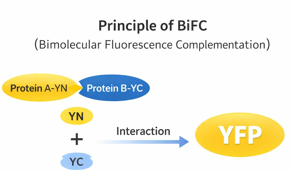
Figure 1. Principle of BiFC assay.
3. Applications of BiFC
BiFC is widely used in plant molecular biology to study protein–protein interactions under near-physiological conditions.
- Identify protein–protein interactions in vivo
- Validate predicted interactions
- Study signaling pathways
- Determine interaction localization
- Analyze plant stress and immune responses
👉 BiFC is especially valuable for confirming interactions directly in living cells.
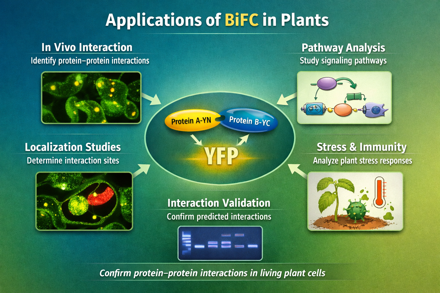
Figure 2. Applications of BiFC.
4. Materials and Reagents
4.1 Biological Materials
- Plant materials (e.g., Nicotiana benthamiana)
- Agrobacterium tumefaciens
4.2 Main Reagents
- BiFC vectors (YN / YC)
- Restriction enzymes
- DNA ligase
- PCR reagents
- Antibiotics
- Infiltration buffer
4.3 Consumables
- Centrifuge tubes
- Pipette tips
- Syringes (needleless)
5. Experimental Procedure
Step 1️⃣ Gene Cloning
- Amplify genes of interest by PCR
- Remove stop codon
Step 2️⃣ Vector Construction
- Clone Protein A into YN vector
- Clone Protein B into YC vector
Step 3️⃣ Transformation
- Transform into Agrobacterium
Step 4️⃣ Co-infiltration
- Mix Agrobacterium strains
- Adjust OD600 to ~0.6–1.0
- Infiltrate tobacco leaves
Step 5️⃣ Expression
- Incubate 48–72 hours
Step 6️⃣ Fluorescence Observation
- Observe YFP signal under confocal microscope
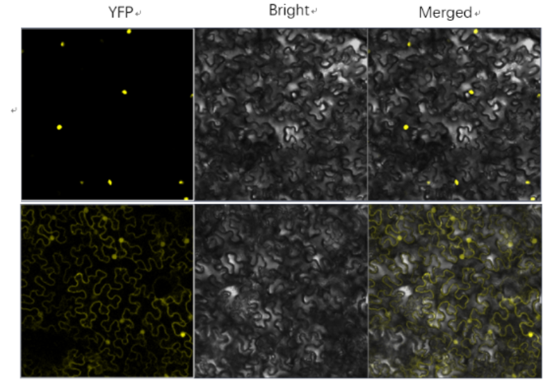
Figure 4. BiFC fluorescence signal indicating protein–protein interaction in plant cells. Strong YFP fluorescence is observed when the two target proteins interact.
6. Data Interpretation
- ✔ Strong fluorescence → strong interaction
- ✔ Weak fluorescence → possible transient interaction
- ❌ No signal → no interaction or expression issue
- 📍 Localization → interaction site
👉 BiFC signals are irreversible once formed.
7. Important Considerations
- ⚠️ Possible false positives
- ⚠️ Fusion orientation matters
- ⚠️ Overexpression effects
- ⚠️ Essential controls:
- Negative control
- Empty vector
- Positive control
8. Conclusion
BiFC is a powerful method for studying protein interactions in plant cells.
- 👉 Direct visualization
- 👉 In vivo validation
- 👉 Spatial information
Need help with your BiFC experiment?
Explore our services: BiFC Analysis Services